
Atrial Fibrillation (A-Fib)
DIEP Flap
Pelvic Fracture
Intramural Hematoma
Osteoporosis
Case Studies
Demographics: 85 y/o male
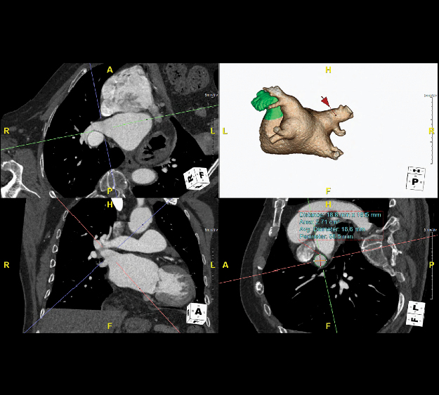
Diagnosis: Persistent atrial fibrillation (A-Fib)
Description of the Condition: Persistent atrial fibrillation is a type of cardiac arrhythmia characterized by irregular and rapid electrical activity in the atria (upper chambers of the heart), and a corresponding heartbeat. The condition is considered persistent once it lasts for more than seven days and requires medical intervention to restore normal heart rhythm.
Description of the 3DLab Work: The 3DLab provided measurements of volume and shape of left atrial appendage (LAA) including depth, width, and ostium measurements, as well as pulmonary veins measurements, including their branches. This type of 3D imaging and measurements help surgeons determine the best “roadmap” for the surgery.
To learn more about A-fib please visit:
https://ucsfhealthcardiology.ucsf.edu/patient-care/clinical-services/electrophysiology-and-arrhythmias/patients/atrial-fibrillation
Image/Video of the 3DLab work:


Demographics: 55 y/o female
Diagnosis: Breast cancer
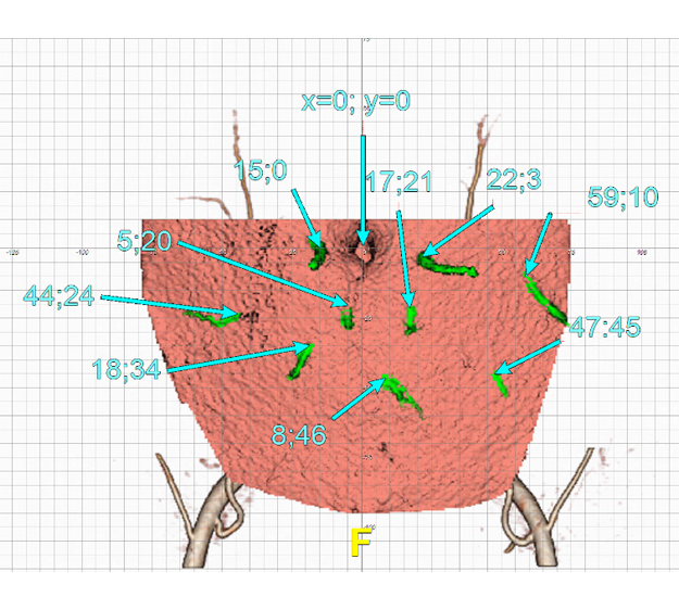
Description of the Condition: Deep Inferior Epigastric Artery Perforator (DIEP) flap breast reconstruction procedure uses a patient's own abdominal skin and fat to reconstruct a breast after mastectomy, reconnecting individual blood vessels to produce a more elastic and natural feel in the breast. Compared to other reconstruction methods, this method better preserves the abdominal muscle and nearby tissue and consequently reduces postoperative pain.
Description of the 3DLab Work: Using state of the art software, the 3DLab performed detailed 3D visualization of DIEP (Deep inferior epigastric perforator) anatomy and measurements for breast reconstruction surgery. This information is used by the surgeons to better plan the procedure and help with resection of the DIEP flap.
To learn more about breast cancer and breast reconstruction procedures please visit:
-
https://cancer.ucsf.edu/breastcarecenter/treatment/surgical_oncology/reconstruction/diep_flap
-
https://surgery.ucsf.edu/conditions--procedures/breast-cancer.aspx
Image/Video of the 3DLab work:


Demographics: female, 65 years old
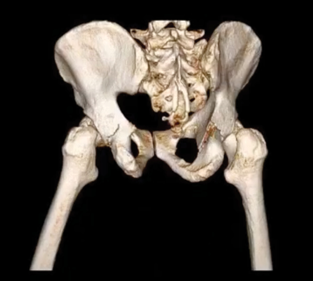
Diagnosis: Pelvic Fracture
Description of the Condition: Pelvic fracture is a break in one or more bones of the pelvis, involving damage to the hip bones, sacrum, or coccyx. They are uncommon, constituting only 3% of all fractures in adults, but may require urgent treatment. In an older person who may have osteoporosis, a lower-impact event such as a minor fall may be enough to cause a pelvic fracture.
Description of the 3DLab Work: The 3DLab provided image reconstruction of left and right coxal (pelvic) bone with subtracted femurs to assist in surgery planning. Isolating only left or only right coxal bone allows the surgeon to see the fracture in 3D view. The Maximum intensity Projection (MIP) enables the surgeon to better understand the morphology and fracture lines.
To learn more about pelvic fractures please visit:
https://orthoinfo.aaos.org/en/diseases--conditions/pelvic-fractures/
Video of the 3DLab work:
Demographics: 85 y/o male
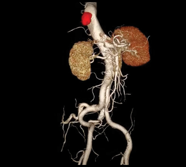
Diagnosis: Thoracic intramural hematoma
Description of the Condition: Thoracic intramural hematoma is a condition where blood accumulates within the layers of the aortic wall, causing vessel layers to separate and create a pocket of blood within the wall itself. The subsequent compression on the aorta can narrow the passageway through which blood flows, potentially leading to reduced oxygen supply to the organs and tissues downstream from the affected area.
Description of the 3DLab Work: The 3DLab provided a 3D reconstruction of aorta showing the hemorrhage and aorta in 360 view. This information can be beneficial for surgeons to identify and understand the position of hemorrhage in relation to aorta and better plan treatment and surgery.
To learn more about Thoracic intramural hematoma please visit:
https://www.youtube.com/watch?v=z7YSNDNHHSY
Image/Video of the 3DLab work:
Demographics: 75 y/o male
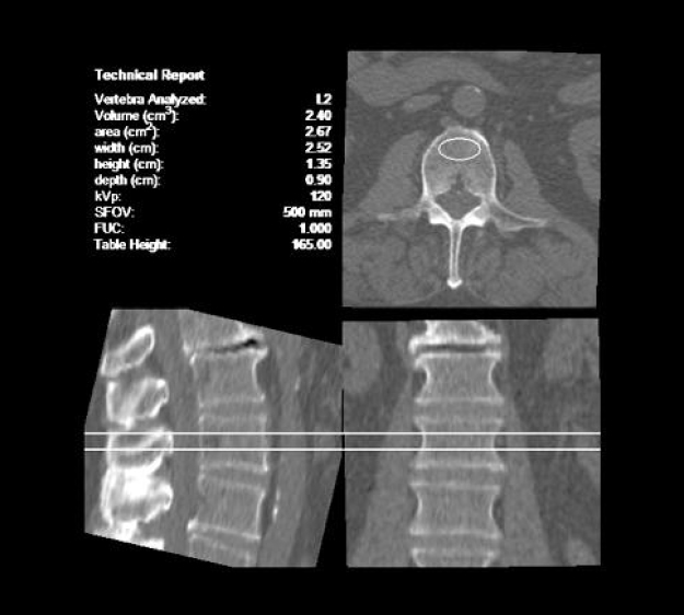
Diagnosis: Osteoporosis
Description of the Condition: Osteoporosis is a progressive bone disease characterized by reduced bone density and increased fragility, leading to an elevated risk of fractures. It occurs when the body loses bone mass faster than it can be replaced. Commonly associated with aging, osteoporosis can be influenced by other factors such as hormonal changes, genetics, and lifestyle choices.
Description of the 3DLab Work: The 3DLab assessed the bone mineral density (BMD) measurement of the spine (L1 to L3) and the proximal femur using CT images and FDA approved volumetric Mindways QCT software. This information is used for diagnosis, assessment of fracture risk, and assessment of treatment response.
To learn more about Osteoporosis please visit:
- www.ucsfhealth.org/conditions/osteoporosis
- www.radiology.ucsf.edu/patient-care/services/osteoporosis
Image/Video of the 3DLab work:


