Projects

Machine and Deep Learning Applied to MR Images to Characterize Degenerative Joint Disease

Principal author

Valentina Pedoia, PhD
Associate Professor

Contributors

Sharmila Majumdar, PhD
co-Executive Director, Science & Technology Resource Group, Scientific Director
Overview

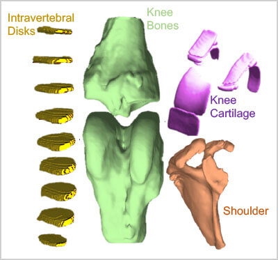
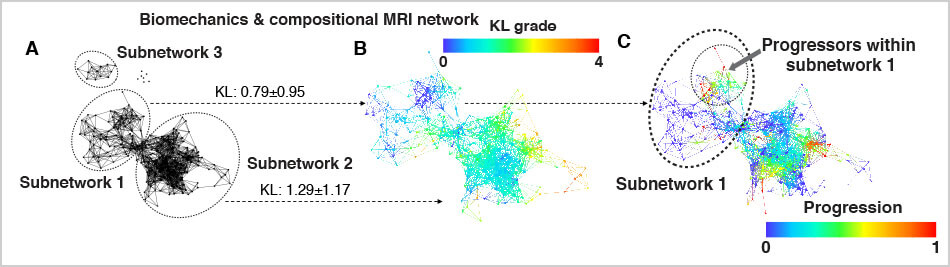
Valentina Pedoia, PhD, and Sharmila Majumdar, PhD, respectively an Assistant Professor and Professor in the Department of Radiology and Biomedical Imaging, are developing algorithms for advanced computer vision and deep learning for improving the usage of non-invasive imaging as diagnostic and prognostic tools of degenerative joint disease. Their lab has developed deep learning convolutional neural networks for musculoskeletal tissue segmentation, abnormality detection, and severity staging covering a diverse range of imaging modalities and diseases – including bone fractures, soft tissue degeneration, and sports injuries. This work has been supported by NIH R00 and R61 grants, as well as the Department’s ongoing partnership with GE Healthcare.

3D modeling from MRI images of different anatomies
Fully Automatic segmentation and 3D modeling from MRI images of different anatomies.
Multi modal graph of a Knee OA
Multi modal/Multisource graph of a Knee OA population (A) Extracted network based on biomechanics and compositional MRI variables showing the presence of three distinct sub-networks marked with dashed circles. (B) The same network is shown colored by Kellgren and Lawrence (KL) grade (OA severity). The combined network of biomechanics and compositional MRI showed differences in osteoarthritis severity between the subnetwork 1 (prevalence o blue nodes low KL grading) and subnetwork 2 (prevalence of red nodes high KL grading). (C) the subjects in the progression cohort are located in both subnetworks. However, In subnetwork 1 progressors are located in a specific region marked with a dashed circle.
Goals
Methodology
Desired clinical outcomes