Clinicians frequently perform magnetic resonance imaging (MRI) of the lumbar spine in patients with lower back pain (LBP), there is a need for standard operating procedures (SOPs) for spinal imaging protocols to better facilitate data integration across studies and sites.
To solve this issue, The Back Pain Consortium (BACPAC) Spine Imaging Working Group developed SOPs for MRI of the lumbar spine. The study, titled "Magnetic resonance imaging of the lumbar spine-recommendations for acquisition and image evaluation from the BACPAC Spine Imaging Working Group," is published in Pain Medicine.
"The SOPs are meant to improve understanding of pain mechanisms and facilitate patient phenotyping by codifying MRI-based methods that provide standardized, non-invasive assessments of spinal pathologies," write the investigators, led by Nico Sollmann, MD, PhD, a UCSF postdoctoral scholar at the UCSF Department of Radiology and Biomedical Imaging.
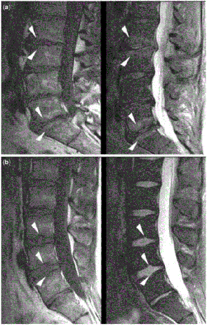
The authors share SOPs for both the imaging protocol — including setup and sequence planning and imaging sequences — and the evaluation of imaging data — including Modic changes, endplate defects, intervertebral disc changes, facet and sacroiliac joint changes and stenosis. These SOPs address two potential sources of variability in MRI for patients with LBP: image acquisition and image interpretation.
Following these SOPs "may help studies and sites better integrate data and perform robust analyses and inferences with much larger datasets than before," says Dr. Sollmann. The SOPs are also "meant to improve understanding of pain mechanisms and facilitate patient phenotyping by codifying MRI-based methods that provide standardized, non-invasive assessments of spinal pathologies," write the investigators.
The BACPAC Spine Imaging Working Group developed this approach for use in BACPAC studies, but the procedures could also be applied at other centers with the same benefits of better harmonized MRI data from the standardization of imaging acquisition and interpretation.
Additional authors from UCSF ci2 include senior author Roland Krug, PhD, Sharmila Majumdar, PhD, Cynthia Chin, MD, Duygu Tosun-Turgut, PhD, Misung Han, PhD, Eugene Ozhinsky, PhD and Thomas Link, MD, PhD.