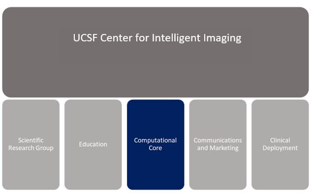
The Computational Core, one of five Pillars[i] within UCSF’s Center for Intelligent Imaging (ci2), creates data science resources and enables Artificial Intelligence (AI)-based research and development to improve health care. Director Jason Crane, PhD leads the specialized team which includes computational data scientists Bruno Astuto-Arouche Nunes, PhD, Pablo Damasceno, PhD, James Hawkins, Beck Olson and Madeline Hess. Rutwik Shah, MD, project director, clinical product innovation and research partnerships, is also a key collaborator. The Core’s Infrastructure subgroup, consisting of Jeff Block, director of infrastructure, Peter Storey, scientific computing services manager, Emma Bahroos, imaging program manager, Wyatt Tellis, PhD, director of innovation and analytics and John Mongan MD,PhD, director of translational informatics and the Clinical Deployment pillar in the Ci2 provide expertise in the computational infrastructure underpinning the center’s resources.
The Computational Core team also includes the 3D Laboratory which functions to provide clinical and research image analysis services. The team includes imaging scientists Vanitha Sankaranarayanan, MS, and Felix Liu, MS, as well as Shezhang Lin, MD, an image analysis and visualization lead, Meng Lian, MS, and Ziba Mansoori, MD clinical imaging IT specialists, Songling Liu, MD, a research data analyst, Bo Fan, MD, a specialist, and Tijana Popovic, MS, a staff research associate.
Researchers across UCSF are encouraged to request the services of the Computational Core for projects that would benefit from a collaborative approach to AI and machine learning-based research. The Core’s services include:
- Building computational infrastructure, data science tools, and platforms
- Developing big data resources
- Providing core competency in machine learning and software development
Current projects
The team produces and manages a range of innovative data science projects in various stages of design, development, and validation at UCSF. The goals for this work are:
1. Translating novel ideas from concept to clinical deployment
2. Using the potential of imaging to impact patient care – proceed from a strategy of fostering AI-based imaging innovation and streamlining clinical validation
Current projects leveraging the team’s expertise include:
- A brain tumor segmentation project aimed at calculating tumor volumes to assess disease progression
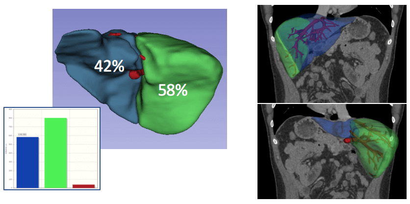
- Automated data analysis for liver transplant surgery planning
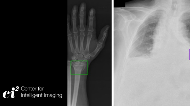
- Work directed at rapidly predicting the likelihood of hip fractures from X-ray images
"Our brain tumor segmentation project will help us tackle one of the hardest problems we face in neuro-oncologic imaging – inter-reader variability in glioma brain tumor follow-up measurements. By automatically segmenting gliomas this tool will provide radiologists and neuro-oncologists with reliable information on tumor size for response assessment that will ultimately lead to better decision making for both standard of care treatments and clinical trials,” says Javier Villanueva-Meyer, MD, neuroradiologist and the project’s clinical PI. “Also exciting is the fact that these segmentations will fuel research by providing expert approved regions of interest that can be readily applied to our myriad ongoing quantitative MRI and molecular imaging projects."
Beck Olson is building a model conceived by Spencer Behr, MD and Z. Jane Wang, MD, ci2 members and faculty in the Abdominal Imaging section of UCSF Radiology. “Currently much patient data is manually delineated to prepare for liver transplants, which can be cumbersome,” says Olson, who was able to drastically decrease the amount of time needed to map an image of a liver using AI.
Sharmila Majumdar, PhD, executive and scientific director of the ci2 leads clinical validation and deployment of the hip fracture project. “In hip fractures, the time to treatment is important,” notes Dr. Majumdar. “In a suspected hip fracture, the patient goes to the emergency room (ER) and has an X-ray, the time to read the X-ray can be variable and long. We have developed a method to evaluate the X-ray, predict the fracture, thus providing rapid results.”
Model Validation
“The prospective validation stage of the work is very important for all projects that are developed and managed by the Computational Core. It’s important to create models that fit into the clinical workflow, that are feasible from an engineering perspective and that can be interpreted effectively in a clinical context,” says Crane. “A key question for our work is: Does the invention provide a usable solution to a clinical problem?”
Model approval and implementation
Once models are developed, validated, and ready for incorporation into routine clinical care, they go through an approval and implementation process. John Mongan, MD, PhD, observes that “effectively integrating an AI algorithm into clinical care is often at least as complex as developing the algorithm in the first place. We’ve developed a framework for evaluating AI algorithms to assess whether they have sufficient quality and performance to merit clinical deployment, guiding the form of that deployment and ensuring longitudinal post-deployment monitoring and QA.”
Industry collaboration
ci2 collaborates with external partners including manufacturers of imaging equipment, AI development companies such as Kheiron and technology companies including NVIDIA. A recent project with NVIDIA involved using federated learning across a 20-site international consortium to develop an AI model to predict COVID outcomes (O2 requirements) from chest X-rays and emergency department data.
“We have a great, highly motivated team,” notes Crane. “Our members are collaborative, smart, and we have a number of exciting new projects underway. We look forward to contributing to big improvements in health care workflow and outcomes for patients.”
[i] The other four ci2 Pillars: Science and Technology Resource Group (SRG), Clinical Deployment, Internships and Education, and Marketing and Communications.

